
Understanding the Difference Between 3-Lead and 5-Lead ECG Monitors
I. Differences Between 3-Lead and 5-Lead ECG Monitors
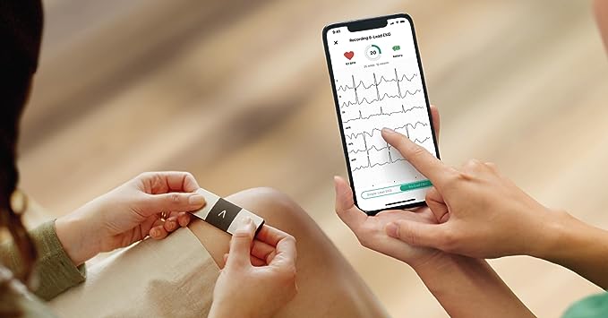
The 3-lead ECG monitor captures ECG readings from three leads: I, II, and III. In contrast, the 5-lead ECG monitor captures readings from five leads: I, II, III, aVR, aVF, aVL, and V.
To facilitate quick connection, electrode pads are color-coded. For the 3-lead ECG, the wires are marked as red, yellow, green (or white), black, and red. For the 5-lead ECG, the wires are marked as white, black, red, green, and brown.
The electrodes’ positions differ between the two systems, even when they share the same color code. Using the abbreviations RA, LA, RL, LL, and C is more reliable than memorizing the colors.
II. Positioning of Three Leads and Five Leads for ECG Monitoring
1. Positioning of Three Leads:
- Right Arm (RA): First intercostal space at the midclavicular line on the right sternal border.
- Left Arm (LA): First intercostal space at the midclavicular line on the left sternal border.
- Left Leg (LL): Level of the xiphoid process on the left midclavicular line.
2. Positioning of Five Leads:
- Right Arm (RA) – White Line: First intercostal space at the midclavicular line on the right sternal border.
- Right Leg (RL) – Green Line: Level of the xiphoid process on the right midclavicular line.
- Chest (C) – Brown Line: Fourth intercostal space on the left side of the sternum.
- Left Arm (LA) – Black Line: First intercostal space at the midclavicular line on the left sternal border.
- Left Leg (LL) – Red Line: Level of the xiphoid process on the left midclavicular line.
III. Operating Points of ECG Monitoring
- Ensure the skin is clean and dry. Use abrasive paper to prepare the skin at the locations where the electrode patches will be placed: below the right clavicle, the intersection of the fifth rib on the left, and the left anterior axillary line, and the midpoint between the nipples.
- Rinse the skin with clean water and dry it with a paper towel or let it dry naturally.
- Prepare the electrode patches.
- Attach the lead wires to the electrode patches: place the red electrode below the right clavicle, the yellow electrode at the midpoint between the nipples, and the black electrode at the intersection of the left fifth rib and the left anterior axillary line.
- Insert the other end of the ECG lead wires into the monitoring device following the arrow direction. Turn on the device to begin ECG monitoring.